-
InLiquid VinylAlcohol Optical Photovoltaics Worcester_Polytechnic_Institute Optoelectonics Fiber Gallium_Arsenide HexagonalBoronNitride ForceDistanceSpectroscopy PhthalocyaninePraseodymium Temasek_Lab neodymium_magnets Conductivity Lanthanum_aluminate Cobalt Pattern StrontiumTitanate Polyaniline Granada Insulator small_scan CHRYSALIS_INC TungstenDeposition Reduction FM-KPFM Heat VerticalPFM Pinpoint PFM IVSpectroscopy Glass Oxidation ConductingPolymer ConductiveAFM Crystal
Report image
If you found this image unacceptable, please let us know. We will review your report and take action if we determine this image is really unacceptable.
ER 316L Stainless Steel
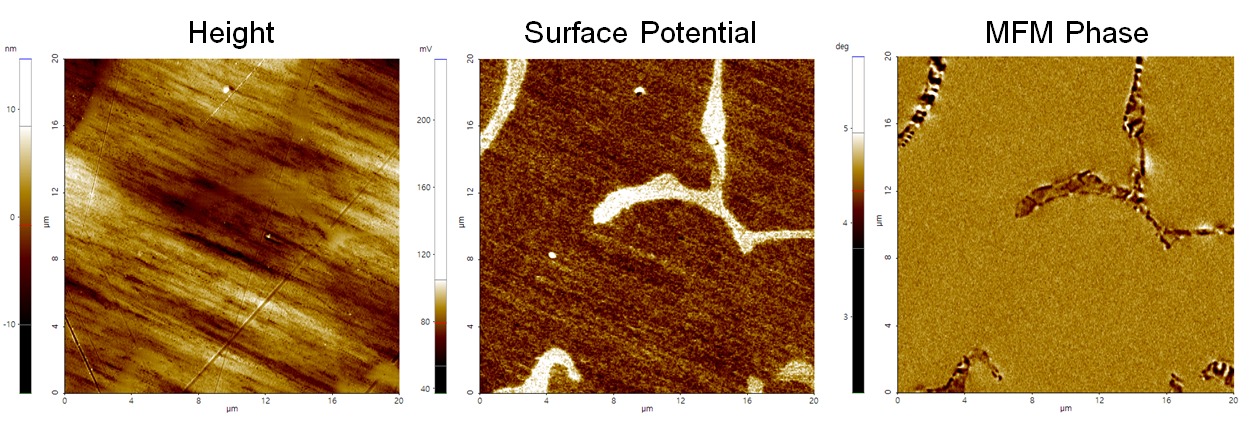
The ferrite MFM phase has a striped appearance due to its ferromagnetic behavior, while the paramagnetic austenite phase shows a uniform appearance.
Scanning Conditions
- System: NX20
- Scan Mode: AM-KPFM, MFM
- Cantilever: PPP-MFMR (k=2.8N/m, f=75kHz)
- Scan Size: 20μm×20μm
- Scan Rate: 0.3Hz
- Pixel Size: 512 × 512
- Lift height: 30nm
- Scan Mode: AM-KPFM, MFM
- Cantilever: PPP-MFMR (k=2.8N/m, f=75kHz)
- Scan Size: 20μm×20μm
- Scan Rate: 0.3Hz
- Pixel Size: 512 × 512
- Lift height: 30nm