-
Heat Wildtype TyphimuriumBiofilm Granada Monisha SFAs ImideMonomer FailureAnlaysis Polarization OpticalModulator electrospinning PinpointNanomechanicalMode Plug Hafnia Dr.JurekSadowski SingleLayer NusEce multi_layer Pipette Tungsten_disulfide BaTiO3 Zhi Barium_titanate LightEmission molecular_self_assembly LightEmiting phase_change TemperatureControl Ucl Protein Implant small_scan IVSpectroscopy UTEM TemperatureControlledAFM
Report image
If you found this image unacceptable, please let us know. We will review your report and take action if we determine this image is really unacceptable.
ER 316L Stainless Steel
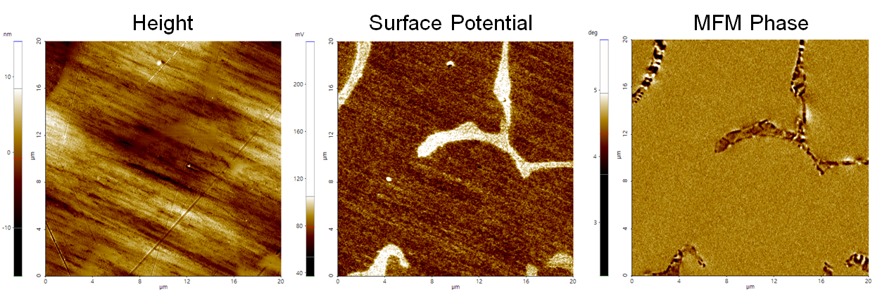
The ferrite MFM phase has a striped appearance due to its ferromagnetic behavior, while the paramagnetic austenite phase shows a uniform appearance.
Scanning Conditions
- System: NX20
- Scan Mode: AM-KPFM, MFM
- Cantilever: PPP-MFMR (k=2.8N/m, f=75kHz)
- Scan Size: 20μm×20μm
- Scan Rate: 0.3Hz
- Pixel Size: 512 × 512
- Lift height: 30nm
- Scan Mode: AM-KPFM, MFM
- Cantilever: PPP-MFMR (k=2.8N/m, f=75kHz)
- Scan Size: 20μm×20μm
- Scan Rate: 0.3Hz
- Pixel Size: 512 × 512
- Lift height: 30nm