SICM Image of Suspended Collagen Fibrils
SICM Image of Suspended Collagen Fibrils
Tatsuo Ushiki and Masato Nakajima (Niigata University, Japan)
 Ion conductance microscopy (SICM) is useful for obtaining contact-free images of the surface topography and has been used for imaging cultured cells under liquid conditions. The Approach-Retract-Scan (ARS) mode is a new mode of SICM, which is especially powerful for imaging samples with steep slopes. To examine the applicability of the ARS mode for imaging biological samples with greater height gaps, dense collagen fibril networks were examined by SICM.
Sample preparation
Collagen fibrils were obtained from the tail tendon of adult Wistar rats and stored in physiological saline with 1-10 % tymol (2-isopropyl-5-methylphenol) at 4 °C for a minimum of 1 day. A small piece of the tendon was then stretched on the surface and air dried overnight, followed by immersion of the sample in physiological saline. SSICM imaging was made in the ARS mode using XE-Bio System
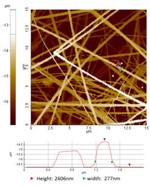
Figure 1 Dense collagen fibril networks imaged by the ARS/hopping SICM mode SICM. Line profile analysis is based on the red line indicated in the SICM image.Conclusion
Figure 1 is an example of SICM imaging of collagen fibril networks using the ARS mode. The width of the individual fibrils varied from 50 to 470 nm. The section profile also showed that the width of the collagen fibril indicated by green arrows was 277 nm and the height difference between the glass surface and the fibril was 2606 nm. This result indicates that some fibrils are suspended over the glass substrate during scanning for SICM imaging. Thus, the ARS mode of SICM has the advantage of minimizing (or free from) the loading force, which is unavoidable in AFM.
Reference
1P. K. Hansma, B. Drake, O. Marti, S. A. Gould, C. B. Prater, Science 243, 641 (1989).
2Y. E. Korchev, C. L. Bashford, M. Milovanovic, I. Vodyanoy, M. J. Lab, Biophys. J. 73, 653 (1997).
3P. Novak, C. Li, A. I. Shevchuk, R. Stepanyan, M. Caldwell, S. Hughes, T. G. Smart,, et al.. Nat. Methods 6, 279 (2009).
4T. Ushiki, M. Nakajima, M. Choi, S.-J. Cho, F. Iwata. Micron (2012, in press)
